

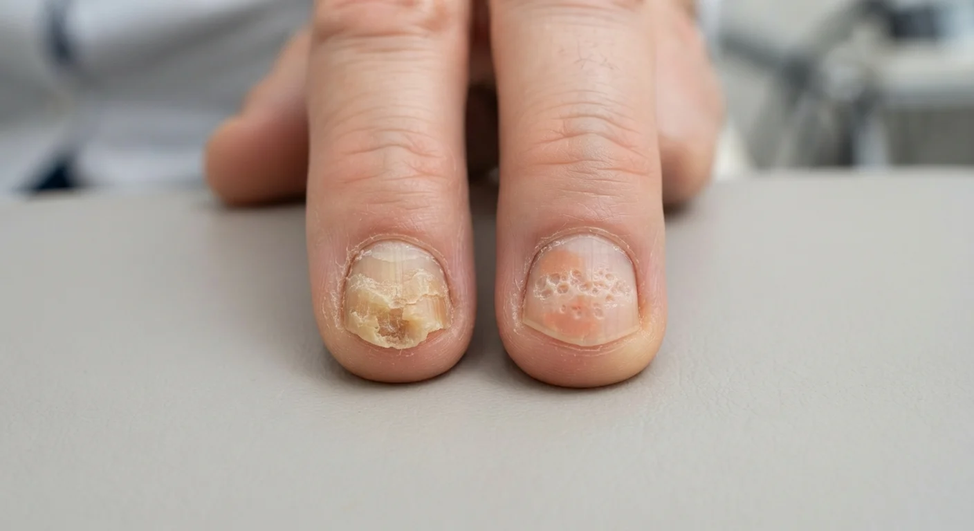
Key Takeaways
Option A
Fungal nail infection (onychomycosis)
An infectious condition caused by dermatophyte or yeast organisms invading the nail.
Best for: Identifying a contagious nail condition that typically starts at the nail's free edge and spreads inward.
Option B
Nail psoriasis
An inflammatory autoimmune condition affecting the nail matrix, bed, or fold.
Best for: Recognizing a non-infectious nail condition usually accompanied by psoriatic changes elsewhere on the skin or joints.
If your nail changes began at the free edge and you frequent shared showers or pools
Fungal nail infection (onychomycosis)
Onychomycosis typically starts at the nail tip or sides and spreads toward the cuticle; exposure to warm, moist communal environments is a well-established risk factor.
If you already have a diagnosis of plaque psoriasis or psoriatic arthritis
Nail psoriasis
Nail involvement occurs in a large proportion of people with skin or joint psoriasis, so nail changes in this context are more likely autoimmune than infectious in origin.
If the affected nail shows small pits or a salmon-coloured spot beneath the nail plate
Nail psoriasis
Pitting and oil-drop (salmon patch) discolouration are hallmarks of nail psoriasis and are not characteristic of fungal infection.
If only one nail is affected and microscopy or culture is available
Fungal nail infection (onychomycosis)
Fungal infection can affect a single nail, and a positive KOH test or fungal culture confirms the diagnosis definitively, ruling out psoriasis.
If nail changes appear alongside joint pain, swelling, or skin plaques
Nail psoriasis
Systemic inflammatory signs point toward psoriatic disease; a dermatologist or rheumatologist should evaluate the full clinical picture.
Why these two conditions are so often confused
Thickened nails, yellow-brown discolouration, and nail plate separation are common to both fungal nail infection and nail psoriasis. Without lab testing, even experienced clinicians can misidentify one for the other. Estimates in dermatology literature suggest that a proportion of nails clinically suspected of fungal infection turn out to be psoriatic on culture, and vice versa. That diagnostic uncertainty matters because antifungal treatment does nothing for an autoimmune condition, and immune-modulating therapies for psoriasis would be unnecessary, and potentially inappropriate, if the cause is actually a living organism.
For general context on how fungal organisms differ from inflammatory conditions, see our overview of fungal infections.
| Criterion | Fungal nail infection | Nail psoriasis |
|---|---|---|
| Cause | Dermatophyte fungi or yeasts | Autoimmune inflammation |
| Contagious | Yes | No |
| Most common site | Toenails | Fingernails (though both can occur) |
| Typical onset location | Distal (free edge) or lateral | Nail matrix or bed; diffuse |
| Nail pitting | Uncommon | Common and characteristic |
| Oil-drop (salmon patch) sign | Absent | Present |
| Subungual debris | Crumbly, powdery material | Hyperkeratotic scaling |
| Associated conditions | Athlete's foot, diabetes, immune suppression | Skin plaques, psoriatic arthritis |
| Diagnostic test | KOH prep, culture, PAS stain | Clinical evaluation, biopsy |
How each condition changes the nail
Fungal nail infection (onychomycosis) typically begins at the distal free edge or the lateral sides of the nail and moves proximally toward the cuticle. The nail plate thickens and becomes brittle, with subungual debris (crumbly material beneath the nail) accumulating as the infection progresses. Colour shifts toward yellow, white, or brown. In severe cases the nail separates from the nail bed, a process called onycholysis.
Nail psoriasis produces a different set of changes driven by immune-mediated inflammation at the nail matrix and nail bed. Pitting (small ice-pick-like depressions on the nail surface) is one of the most specific signs and results from abnormal matrix cell turnover. An oil-drop or salmon-patch sign, a yellow-red discolouration visible through the nail plate, reflects inflammatory changes in the nail bed. Onycholysis also occurs in psoriasis, but the separated area often has a characteristic erythematous (red) border that is less common in fungal cases. Splinter haemorrhages and thickening of the skin beneath the nail (subungual hyperkeratosis) appear in both conditions, which is one reason overlap is so common clinically.
One practical distinction: psoriasis frequently affects the fingernails, whereas onychomycosis is more prevalent in toenails, though neither pattern is absolute.
Context clues that point toward each diagnosis
Pattern and context give clinicians and patients useful early signals, even before testing.
Factors that suggest fungal infection include: a history of athlete's foot (tinea pedis) on the same foot, use of communal showers or pools, diabetes or immune suppression (both raise fungal susceptibility), and a slow progression starting at the nail tip. Certain systemic conditions meaningfully increase fungal risk, and nail infection sometimes follows untreated skin fungus on adjacent skin.
Factors that suggest nail psoriasis include: a personal or family history of plaque psoriasis, concurrent joint pain (psoriatic arthritis affects roughly 30% of people with psoriasis according to the American Academy of Dermatology), visible skin plaques elsewhere on the body, and nail pitting without the crumbly subungual debris typical of fungal cases.
Importantly, the two conditions can coexist. People with psoriasis have an elevated risk of developing onychomycosis, possibly because psoriatic nail changes create a structurally compromised surface that organisms can colonise more easily. A positive fungal culture in someone with known psoriasis does not rule out concurrent psoriatic nail involvement.
Both conditions can occur at the same time
Having nail psoriasis does not protect against fungal infection. Psoriatic nail changes can structurally weaken the nail plate, making colonisation by dermatophytes easier. If a person with known psoriasis notices a sudden worsening of nail appearance, a fungal culture is worth discussing with their dermatologist to check for a secondary infection rather than attributing all changes to the autoimmune condition alone.
Getting a definitive answer
A dermatologist will typically use one or more objective tests before committing to a treatment plan. A potassium hydroxide (KOH) preparation involves dissolving nail clippings in KOH solution so that fungal hyphae (thread-like structures) become visible under a microscope. Fungal culture takes longer (weeks) but identifies the specific organism. Nail biopsy with periodic acid-Schiff (PAS) staining is the most sensitive method for detecting fungal elements and can simultaneously show psoriatic histological features if present.
Psoriasis has no single confirmatory lab test for nail involvement. Diagnosis rests on clinical findings, the broader skin and joint picture, and ruling out infectious causes.
If you are seeing nail changes that concern you, a dermatology consultation is the appropriate next step rather than self-treatment, particularly because oral antifungal medications carry potential drug interactions and are not appropriate for everyone. For broader hygiene practices that may reduce fungal exposure while you await evaluation, practical hygiene guidance is available here.
This article is for general informational purposes only and is not a substitute for professional medical advice, diagnosis, or treatment. Consult a qualified healthcare provider with any questions about your nail health or symptoms.