

Key Takeaways
Muscle repair and hypertrophy
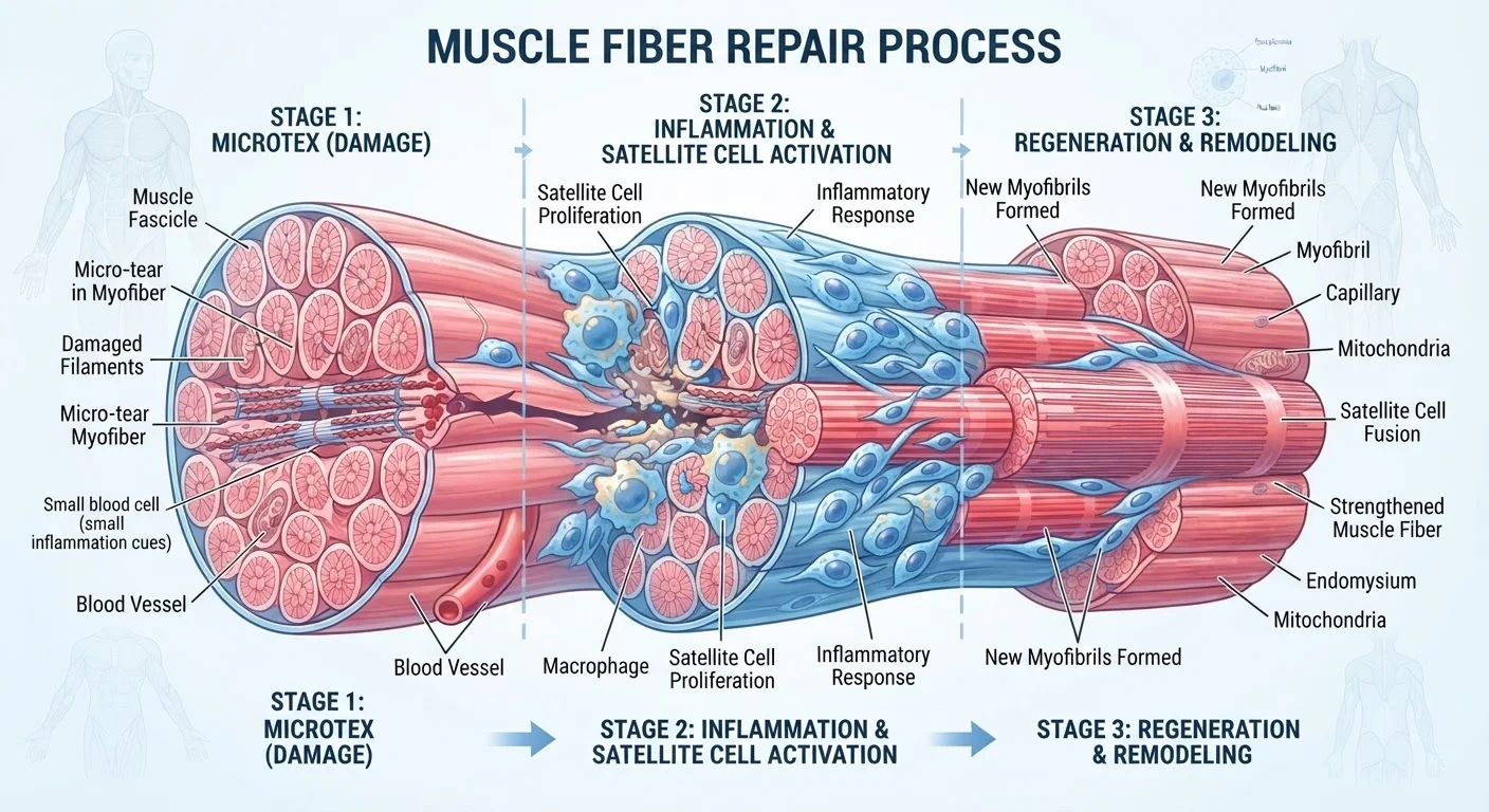
When you exercise, microscopic damage occurs inside muscle fibers. The body responds by clearing damaged tissue, recruiting specialized cells to rebuild, and synthesizing new proteins that make the repaired fiber slightly thicker and stronger than before. This cycle, repeated over time, is what produces measurable gains in strength and muscle mass.
The net outcome depends on the balance between muscle protein synthesis (MPS) and muscle protein breakdown (MPB). Hypertrophy occurs when MPS consistently exceeds MPB during recovery windows.
What actually tears during exercise
The term 'micro-tear' refers to disruption at the level of the sarcomere, the smallest functional unit inside a muscle fiber. During eccentric contractions (the lowering phase of a bicep curl, for example, or the downhill portion of a run), the fiber is lengthening while generating force. That combination produces mechanical strain that pulls apart the thin and thick protein filaments, actin and myosin, that give muscle its structure.
These disruptions are not random breaks. They follow predictable patterns at the Z-disc, the boundary structure between sarcomeres. Electron microscopy studies show that Z-disc streaming, where the disc broadens and distorts, is one of the earliest signs of exercise-induced muscle damage. The body immediately recognizes this structural change as a signal to begin repair.
For context on where this fits within the broader recovery picture, see what happens inside your body during recovery.
The inflammatory phase: necessary, not harmful
Within minutes of micro-damage, mast cells in the surrounding connective tissue release histamine and other signaling molecules. This triggers vasodilation, widening of local blood vessels, which allows neutrophils (a type of white blood cell) to flood the damaged area within the first few hours. Neutrophils clear cellular debris and release cytokines that recruit macrophages to continue the cleanup process.
This acute inflammation is often misunderstood. Routinely suppressing it with anti-inflammatory medications during recovery may blunt the repair signal. Research published in peer-reviewed sports medicine journals has raised questions about whether habitual non-steroidal anti-inflammatory drug (NSAID) use around exercise sessions interferes with the satellite cell response. Consult a healthcare professional before using any medication to manage exercise soreness.
Let inflammation do its job
Reaching for an ice pack or an over-the-counter anti-inflammatory immediately after every training session may feel prudent, but the acute inflammatory phase is part of the repair signal. Discuss any habitual use of anti-inflammatories around training with a healthcare provider, particularly if you are working toward muscle strength or hypertrophy goals.
By roughly 24 to 48 hours post-exercise, macrophages shift from a pro-inflammatory to an anti-inflammatory profile. This transition coordinates the handoff from debris clearance to active tissue building.
Satellite cells and the rebuilding process
Satellite cells are muscle-specific stem cells that sit dormant between the fiber membrane and the surrounding basal lamina. Mechanical stress and the cytokine signals released during inflammation activate them. Once active, satellite cells proliferate and fuse with the damaged fiber, donating their nuclei to the repair site.
This addition of new nuclei matters because each nucleus in a muscle fiber governs a fixed volume of cytoplasm (the intracellular fluid and organelles). More nuclei mean more sites where protein synthesis machinery can operate simultaneously, which is one mechanism through which repeated training sessions produce larger fibers over time.
48-72 hrs
Typical window for basic fiber repair after moderate exercise
Based on biopsy and ultrasound studies tracking structural muscle damage resolution in healthy adults.
Up to 48 hrs
Duration of elevated muscle protein synthesis post-exercise
Observed in untrained individuals; the window shortens as training adaptation progresses, per exercise physiology research.
Less than 7 hrs
Sleep duration linked to reduced anabolic signaling
Multiple observational studies associate consistently short sleep with lower testosterone and growth hormone output in adults.
The satellite cell response is also sensitive to nutrition and sleep. Growth hormone, released mainly during slow-wave sleep, promotes insulin-like growth factor 1 (IGF-1), which directly stimulates satellite cell proliferation. Inadequate sleep therefore affects repair at a cellular level, not just at the level of perceived fatigue.
Protein synthesis: the construction phase
Muscle protein synthesis (MPS) is the process by which ribosomes inside muscle cells assemble amino acids into new contractile proteins. Exercise elevates MPS within two to four hours of a session, and this elevated rate can persist for 24 to 48 hours in untrained individuals or for a shorter window in highly trained athletes whose bodies have adapted to the stimulus.
Dietary protein supplies the amino acids that fuel this process. Leucine, one of the essential branched-chain amino acids, has a well-documented role as a direct trigger for MPS via the mTOR (mechanistic target of rapamycin) signaling pathway. This is why the timing and quality of protein intake after exercise is a legitimate practical concern rather than a marketing construct. For a detailed breakdown, the recovery nutrition guide covers protein timing and carbohydrate replenishment strategies.
The liver also contributes to this process by synthesizing albumin and other plasma proteins that support tissue repair and nutrient transport. For readers interested in metabolic health more broadly, the liver health hub provides relevant background.
How recovery methods support the repair cycle
Sleep, nutrition, and movement intensity are the three variables with the most consistent support in the research literature. Sleep protects the anabolic window when growth hormone peaks. Protein intake supplies the amino acid substrate for MPS. Managing training volume prevents the repair process from being interrupted before it completes.
Active recovery (low-intensity movement on rest days) can increase local blood flow, which may assist in clearing inflammatory metabolites without adding new mechanical stress. The comparison of active recovery and complete rest examines how to choose between these approaches based on training context. For those interested in practical movement strategies that reduce physical discomfort, the mobility and recovery hub is a useful reference.
Recovery capacity is not fixed. It changes with age, stress load, sleep quality, and training history. The recovery across the lifespan article addresses how these variables interact over decades. For a comprehensive framework that integrates all these factors, see building a whole-body recovery framework.
This article is for informational purposes only and is not medical advice. Consult a qualified healthcare professional for guidance specific to your health history and recovery needs.