

Key Takeaways
Hair follicle miniaturisation
Hair follicle miniaturisation is the gradual shrinking of individual hair follicles over successive growth cycles, producing progressively thinner and shorter strands until the follicle can no longer support visible hair. It is the biological process at the root of pattern hair loss in both men and women. The follicle does not die outright; it becomes dormant, which is why many cases respond to treatment if addressed early.
Miniaturisation is driven largely by sensitivity to dihydrotestosterone (DHT), a hormone derived from testosterone via the enzyme 5-alpha reductase, which shortens the anagen (growth) phase with each cycle.
The four phases of the hair growth cycle
Every strand of hair on your scalp follows a predictable biological program. The cycle has four phases, and the proportion of follicles in each phase at any given time determines how full your hair appears.
Anagen is the active growth phase, lasting roughly two to seven years depending on genetics and overall health. During anagen, cells in the follicle's dermal papilla divide rapidly, pushing the hair shaft upward. Approximately 85 to 90 percent of scalp follicles are in anagen at any moment.
Catagen is a brief transition phase lasting about two weeks. Cell division stops, the follicle shrinks, and the hair detaches from its blood supply.
Telogen is a resting phase lasting two to three months, during which the hair is retained in the follicle but no longer growing. About 10 to 15 percent of follicles are in telogen at any point.
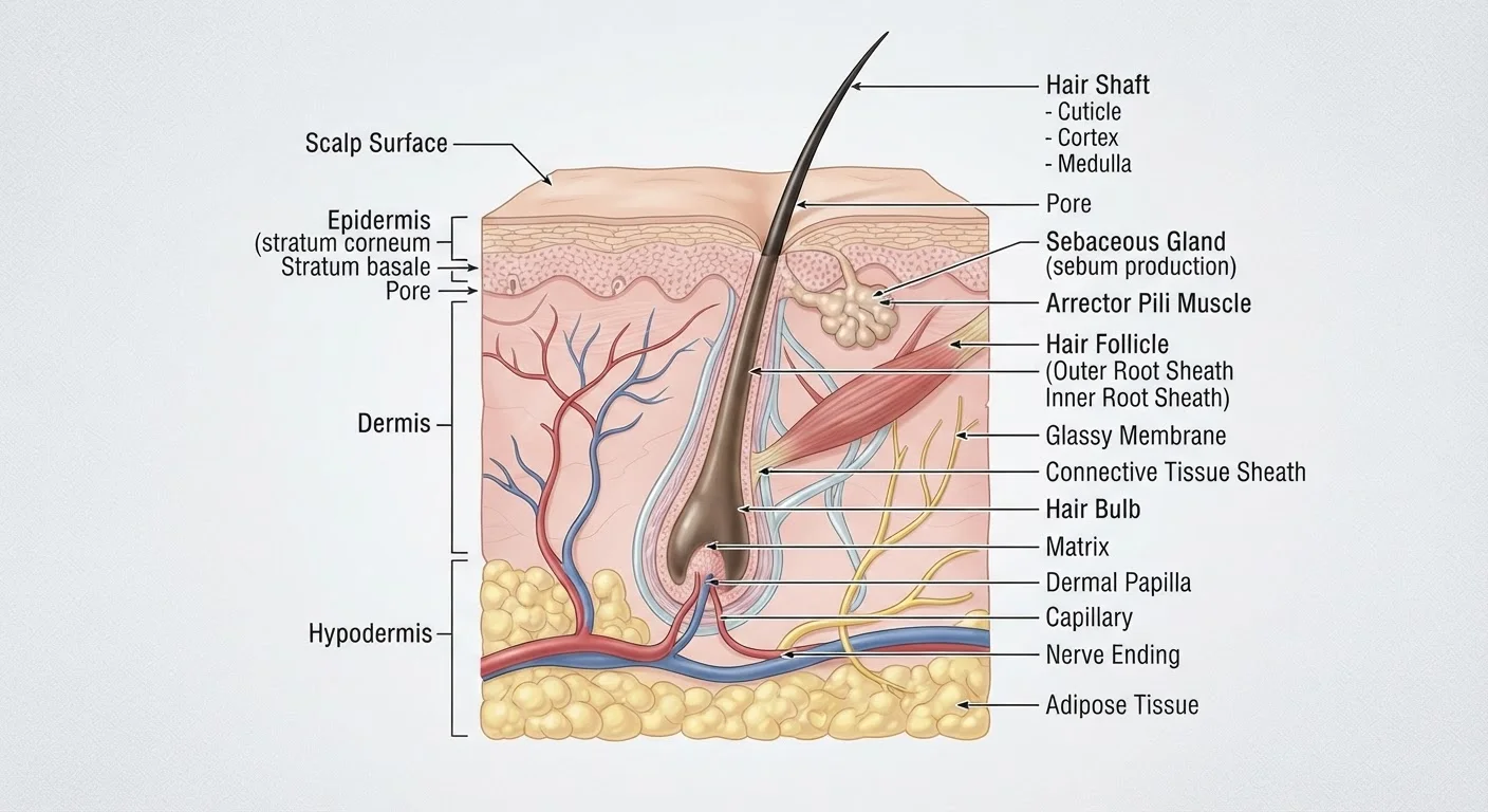
Exogen is when the resting hair is shed, often accelerated by mechanical action like washing or brushing, and a new anagen cycle begins. See our introduction to scalp anatomy for a broader look at follicle structure beneath the surface.
What miniaturisation actually means
Pattern hair loss is not caused by follicles switching off permanently. It is caused by each successive growth cycle producing a shorter anagen phase, so the new hair shaft is thinner and shorter than the one before it. Over several cycles, a terminal hair (thick, pigmented, long) becomes a vellus hair (fine, nearly colorless, short). This process is miniaturisation.
DHT, formed when the enzyme 5-alpha reductase converts testosterone, binds to androgen receptors in genetically susceptible follicles. This binding compresses the anagen phase from years to months. The follicle does not disappear; it persists in a compressed state, which is why follicle counts on biopsy often remain relatively normal even in visible thinning.
Monitor your scalp before thinning becomes visible
Because miniaturisation progresses gradually, many people do not notice significant change until 30 to 50 percent of follicle diameter is already reduced. A dermatologist can use trichoscopy, a non-invasive scalp imaging technique, to detect early follicle changes before they become visible to the naked eye. Earlier assessment generally means more options for intervention.
Because miniaturisation progresses gradually, many people do not notice significant change until 30 to 50 percent of follicle diameter is already reduced. This biological reality reinforces why scalp health monitoring matters even before visible thinning becomes obvious.
How inflammation and blood flow affect follicle function
Follicles are metabolically demanding structures. They require a continuous supply of oxygen, glucose, and micronutrients delivered through the dermal papilla's capillary network. When scalp circulation is poor, follicles in anagen do not receive adequate resources to sustain full growth cycles.
Chronic low-grade inflammation compounds the problem. Inflammatory cytokines can accelerate the catagen transition, pushing follicles out of the growth phase prematurely. Conditions like seborrheic dermatitis and scalp psoriasis create a hostile microenvironment around the follicle opening, and over time this can contribute to thinning even in the absence of androgenetic factors. The connection between scalp health and hair growth covers this relationship in more depth.
The stress pathway and telogen effluvium
Physiological stress, whether from illness, caloric restriction, surgery, or chronic psychological strain, activates the hypothalamic-pituitary-adrenal axis and raises cortisol. Elevated cortisol disrupts the signaling that keeps follicles in anagen. The result is a mass shift of follicles into telogen, followed two to three months later by a wave of shedding called telogen effluvium.
The delay between the stressor and the visible shedding often confuses people: they may not connect hair loss in month three to a high-stress event in month one. Telogen effluvium is usually reversible once the trigger is resolved, but repeated cycles of stress-induced shedding can interact with underlying miniaturisation to accelerate visible thinning. The biological pathway linking cortisol to the hair follicle explains this mechanism in detail.
Nutrition, lifestyle, and follicle biology
Follicle cells are among the fastest-dividing cells in the body, making them sensitive to nutritional shortfalls. Iron, zinc, biotin, and adequate protein all support the enzymatic and structural processes of hair shaft production. Deficiency in any of these can shorten anagen or increase telogen shedding without any hormonal component.
Sleep quality affects circadian rhythm-regulated gene expression in follicle stem cells. Poor sleep alters the timing of growth phase transitions. Physical activity supports dermal circulation, which benefits follicle perfusion. These factors do not replace medical treatment when hormonal or pathological causes are present, but they create the conditions under which follicles can function optimally. The lifestyle factors most consistently linked to follicle health covers the evidence behind these habits.
This article is for informational purposes only and is not medical advice. Consult a qualified healthcare professional for personal concerns about hair loss, scalp conditions, or any changes in health.