

Key Takeaways
DHT (Dihydrotestosterone)
Dihydrotestosterone, commonly abbreviated as DHT, is a hormone derived from testosterone through a conversion process driven by an enzyme called 5-alpha reductase. In genetically susceptible hair follicles, DHT binds to androgen receptors and triggers a process called miniaturization, where the follicle progressively shrinks over successive growth cycles. This is the primary biological driver behind androgenetic alopecia, the most common form of pattern hair loss in both men and women.
DHT has roughly three to five times the androgenic potency of testosterone at the androgen receptor, which explains why even relatively modest DHT levels can produce significant follicular effects in sensitive scalp regions.
How DHT forms and where it acts
Testosterone circulates in the bloodstream in both men and women, though at different concentrations. When testosterone enters certain cells, particularly those in the skin, prostate, and hair follicles, the enzyme 5-alpha reductase converts it into DHT. This conversion happens locally within the tissue, meaning scalp follicles can produce DHT from circulating testosterone without a spike in blood DHT levels being required.
DHT then binds to androgen receptors inside the follicle's dermal papilla cells, the structures at the base of each follicle that control hair growth signals. In follicles that carry a genetic predisposition to androgen sensitivity, this binding triggers a cascade of gene expression changes that disrupts normal growth regulation. For a fuller picture of how follicles are structured and how they cycle through growth phases, see scalp biology fundamentals.
The miniaturization process explained
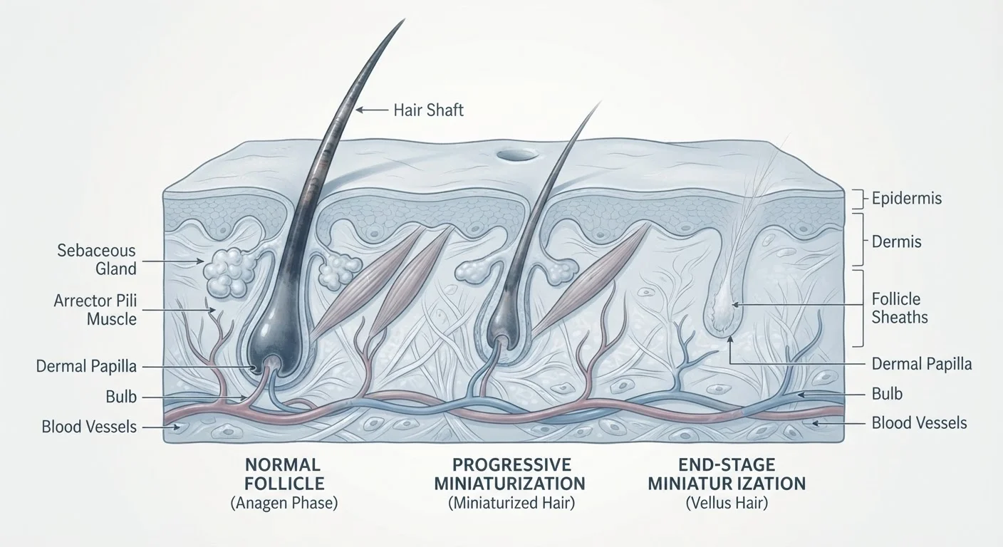
Healthy hair follicles cycle through three phases: anagen (active growth), catagen (transition), and telogen (rest and shedding). In follicles sensitive to DHT, each successive anagen phase becomes shorter. The follicle also physically shrinks with each cycle, producing a thinner, shorter, more lightly pigmented hair shaft. Over years, this progresses until the follicle generates only fine vellus hairs, the near-invisible strands associated with visible baldness.
The mechanism is not a sudden loss event. It is a gradual structural change driven by DHT's interference with growth-promoting signals inside the dermal papilla. Research has identified that DHT suppresses the production of growth factors that normally sustain the anagen phase, while amplifying signals that push follicles into catagen prematurely.
95%
Of pattern hair loss cases attributable to androgens
Androgenetic alopecia is estimated to account for approximately 95% of hair loss in men, according to dermatology literature summarizing prevalence data.
50%
Of men affected by pattern hair loss by age 50
Roughly half of men show some degree of androgenetic alopecia by their fifties, with prevalence increasing with age, as reported across multiple epidemiological studies.
3-5x
Greater androgenic potency of DHT vs. testosterone
DHT binds androgen receptors with substantially higher affinity than testosterone, explaining its outsized effect on androgen-sensitive tissues despite lower circulating concentrations.
Understanding this process matters because it explains why pattern hair loss appears different from other types of shedding. Androgenetic alopecia versus telogen effluvium covers how to distinguish between these two distinct hair loss mechanisms, which have different timelines and causes.
Why genetics determines vulnerability
DHT levels alone do not determine who loses hair. Two people with identical circulating DHT concentrations can have very different hair outcomes depending on how many androgen receptors their follicles express and how sensitive those receptors are to DHT binding. This androgen receptor sensitivity is largely hereditary, encoded in genes that can be inherited from either parent.
The gene variant most consistently linked to male pattern baldness involves the androgen receptor gene on the X chromosome, but inheritance is complex and polygenic. Multiple genes across several chromosomes contribute to overall risk. This is why family history is a useful indicator but not a precise predictor of whether or when someone will develop pattern thinning.
Follicle vulnerability also varies by scalp region. The frontal and crown regions carry higher androgen receptor densities than the occipital (back) region, which explains the characteristic distribution of androgenetic alopecia. This regional difference is also why hair transplants can use donor hair from the back of the scalp: those follicles retain their DHT resistance even after relocation. See the biology of hair loss for more on how follicle miniaturization fits into the broader picture of hair thinning.
DHT in women and hormonal context
Women produce androgens through the ovaries and adrenal glands, and 5-alpha reductase activity in scalp follicles converts some of that testosterone to DHT. In most women, estrogen exerts a counterbalancing effect on androgen signaling, which is one reason female pattern hair loss typically appears later in life and presents differently than male pattern baldness.
During perimenopause and menopause, estrogen levels fall while androgens remain relatively stable or become proportionally more dominant. This hormonal shift can unmask or accelerate DHT-driven follicle sensitivity. Conditions such as polycystic ovary syndrome (PCOS), which involves elevated androgens, are also associated with increased risk of androgen-driven hair thinning in women. Hormonal changes across life stages explores these transitions in depth.
The liver also has a role in androgen metabolism: it processes and clears sex hormones from circulation. Impaired liver function can affect how androgens, including testosterone, are metabolized and cleared, which may in turn influence androgen availability at the follicle level. This connection is covered in the broader context of liver health and metabolic function.
This article is for informational purposes only and is not medical advice. Consult a qualified healthcare provider for guidance on any personal hair loss concerns or before starting any treatment.