
Key Facts About Hormonal Testing: What the Numbers Can and Cannot Tell You

| Most common sample types | Serum (blood), urine, saliva |
| TSH normal reference range (typical adult) | Approximately 0.4 to 4.0 mIU/L (Ranges vary by laboratory and clinical guideline) |
| Total testosterone (adult male reference) | Approximately 300 to 1000 ng/dL (American Urological Association, 2018) |
| Cortisol peak timing | Typically 6 to 8 a.m. (circadian rhythm) |
| Progesterone test timing (luteal phase check) | Day 21 of a 28-day cycle |
| Key confounder for thyroid assays | High-dose biotin supplementation can falsify results (FDA safety communication on biotin interference) |
| Free vs. total testosterone | Free fraction (1 to 3%) is the biologically active portion |
What hormonal tests actually measure
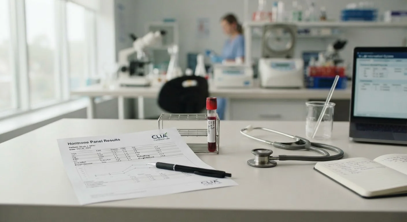
Hormonal testing measures the concentration of specific hormones in a biological sample, typically blood, saliva, or urine. Each method captures a different biological snapshot. A serum blood draw measures the total hormone circulating in plasma at a single moment. Urine panels, especially 24-hour collections, reflect cumulative output over time. Saliva tests are sometimes used for cortisol and certain sex hormones, though their clinical validation varies by hormone and laboratory.
The most commonly ordered hormones include cortisol, thyroid-stimulating hormone (TSH), free thyroxine (T4), free triiodothyronine (T3), estradiol, progesterone, testosterone (total and free), dehydroepiandrosterone sulfate (DHEA-S), luteinizing hormone (LH), and follicle-stimulating hormone (FSH). Each has a distinct physiological role, and no single test captures how the entire endocrine system is functioning. For a broader picture of how these hormones interact, see what hormonal balance means physiologically.
| Most common sample types | Serum (blood), urine, saliva |
| TSH normal reference range (typical adult) | Approximately 0.4 to 4.0 mIU/L (Ranges vary by laboratory and clinical guideline) |
| Total testosterone (adult male reference) | Approximately 300 to 1000 ng/dL (American Urological Association, 2018) |
| Cortisol peak timing | Typically 6 to 8 a.m. (circadian rhythm) |
| Progesterone test timing (luteal phase check) | Day 21 of a 28-day cycle |
| Key confounder for thyroid assays | High-dose biotin supplementation can falsify results (FDA safety communication on biotin interference) |
| Free vs. total testosterone | Free fraction (1 to 3%) is the biologically active portion |
Why reference ranges are not a definitive answer
Every lab report includes a reference range, the interval within which most healthy individuals fall. These ranges are typically derived from population studies, and they reflect statistical norms rather than personal optima. A result that lands within range does not automatically mean that level is appropriate for a specific individual, and a result slightly outside range does not always signal pathology.
Several variables shift where a hormone should ideally sit: age, sex, menstrual cycle phase, time of day, fasting status, recent stress, and the type of assay the laboratory uses. Cortisol, for example, follows a strong circadian rhythm, with levels highest in the early morning and lowest at night. Drawing a cortisol sample at noon versus at 8 a.m. produces meaningfully different numbers, yet both may fall within a broad reference range.
Free versus total hormone measurements add another layer. Total testosterone measures both bound and unbound fractions. Free testosterone, the portion not attached to sex hormone-binding globulin (SHBG) or albumin, is the biologically active fraction. Two people can share identical total testosterone readings while having very different free testosterone levels if their SHBG concentrations differ. This distinction is clinically significant, particularly in older adults. The interpretation challenge is similar in other lab panels; how lipid panel numbers require contextual reading illustrates the same principle in cardiovascular risk assessment.
Common limitations clinicians account for
Hormonal tests are point-in-time measurements of a dynamic system. Pulsatile hormones such as LH are secreted in bursts, meaning a single draw may catch a peak or a trough and give a skewed impression. Reproductive hormones fluctuate across the menstrual cycle, so testing estradiol or progesterone without recording the cycle day makes the number hard to interpret. Clinicians often order tests on specific cycle days, for example day 3 for baseline estrogen and FSH, and day 21 for progesterone in a standard 28-day cycle.
Specimen handling also matters. Delayed centrifugation, improper storage temperature, or a hemolyzed sample can alter measured values. Immunoassay interference from medications, supplements, or biotin (vitamin B7) supplementation has been documented to produce falsely elevated or falsely suppressed thyroid and other hormone readings. Reporting this to the ordering clinician before the blood draw is important.
At-home hormone test kits have grown in availability, and they can provide a starting point for conversation with a clinician. However, their methodologies and quality-control standards vary considerably. The same interpretive caution applies to at-home microbiome testing, as explored in what microbiome test numbers can and cannot tell you.
Symptoms remain essential context. A patient with textbook-range TSH who experiences fatigue, cold intolerance, and hair thinning warrants further evaluation, not reassurance based solely on the number. Lab results are one input in a clinical picture that includes history, physical examination, and symptom patterns. For a structured way to document those symptoms before a clinical visit, this self-assessment checklist can be a useful preparatory tool.
Practical context for reading your own results
When reviewing a hormonal lab report, note the units alongside each value. Testosterone may be reported in ng/dL or nmol/L depending on the laboratory; the numbers look very different, and converting between them requires a fixed factor. Confirm the units before drawing any conclusions.
Hormones do not act in isolation. Estrogen and testosterone both circulate in all adults and influence each other through shared enzymatic pathways. Interpreting estradiol without considering testosterone, SHBG, or progesterone misses meaningful interactions. How estrogen and testosterone share and divide their roles provides useful background for understanding why panels are more informative than individual values.
Liver function also connects to hormonal metabolism. The liver is the primary site for hormone conjugation and clearance, so impaired liver function can alter circulating hormone levels independently of endocrine gland output. Reading liver function tests alongside hormonal panels adds useful context for anyone whose hormonal results look unexpectedly abnormal.
This article is for general informational purposes only and does not constitute medical advice. Always consult a qualified healthcare professional before making decisions about hormonal testing, interpreting results, or changing any aspect of your health care.